50× Micro Endodontic Microscopes: The Science of Seeing What Others Miss
- kairav domadia
- Jan 27
- 4 min read

50× Micro Endodontic Microscopes: Why “Seeing” Is the Real Superpower in Dentistry

You’d be surprised how many dental problems are not about pain—they’re about visibility. A patient walks into Dr Deval Mehta™ with a familiar story: “Doctor, it hurts when I chew… but my X-ray says everything is normal.” They’ve already tried painkillers, avoided that side, and maybe even been told, “We might have to remove the tooth.”
And here’s the truth: many teeth aren’t failing because they’re beyond saving. They’re failing because the problem is too small to see with the naked eye.
That’s exactly where 50× Micro Endodontic Microscopes change the game. They don’t just “zoom in.” They reveal a hidden world—tiny cracks, extra canals, calcifications, missed infections—things that decide whether a tooth gets saved or sacrificed.
Let’s break down the science behind these microscopes and why they’re one of the biggest upgrades modern root canal care has ever received.
What Exactly Are 50× Micro Endodontic Microscopes?
50× Micro Endodontic Microscopes are high-powered dental operating microscopes designed specifically for precision work inside the tooth—especially during root canal treatment and retreatment.
Unlike normal dental loupes (magnifying glasses dentists wear), a microscope provides:
Stronger magnification (up to ~50× in many clinical setups)
Brighter coaxial illumination (light aligned with vision)
A stable, focused field of view for micro-level accuracy
Better depth perception and fine control
Think of it like comparing a basic phone camera to a professional DSLR with studio lighting. Same subject, completely different clarity.
Why 50× Magnification Matters More Than You Think
Your tooth is not a simple hollow structure. Inside it is a complex network of canals—often narrow, curved, branching, or even hidden behind calcified tissue.
At lower magnification, the dentist may still do good work. But at high magnification, dentistry becomes more predictable because the dentist isn’t guessing.
Here’s what 50× Micro Endodontic Microscopes can help reveal:
1) Extra Canals That Regular Eyes Miss
Many teeth—especially molars—can have additional canals that don’t show clearly on standard X-rays. Missing even one tiny canal can allow bacteria to remain, causing pain or reinfection later.
With a microscope, the clinician can locate subtle canal openings, color changes on the pulp floor, and fine anatomical grooves that point to hidden pathways.
2) Hairline Cracks That Decide a Tooth’s Fate
Some cracks are so fine they don’t show up on X-rays and are invisible in routine exams—yet they cause sharp pain while chewing or sensitivity to cold.
Under microscopic magnification and strong illumination, these cracks become easier to detect early—often before the tooth splits or the infection becomes severe.
3) Calcified Canals (The “Blocked Road” Problem)
With age, trauma, or long-standing decay, canals may become calcified—meaning they narrow and harden like a blocked pipeline.
Microscope-assisted micro endodontics helps the dentist safely locate and negotiate these canals with minimal removal of healthy tooth structure—protecting the tooth’s strength.
The Science of Light: Why Illumination Is Half the Magic
Magnification without light is like trying to read tiny print in a dark room. The most powerful advantage of microscopes is not only zoom—it’s coaxial illumination.
Coaxial illumination means the light beam is aligned with the dentist’s line of sight, so shadows are reduced and deep areas inside the tooth become clearly visible. This is critical when working in tight spaces where traditional dental chair lights can’t reach effectively.
That’s why 50× Micro Endodontic Microscopes aren’t just “bigger lenses.” They are an entire visibility system.
Micro Endodontics vs Traditional Root Canal: What Changes in Real Life?
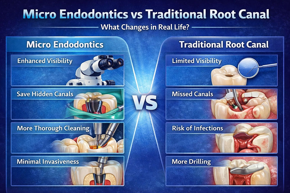
A microscope doesn’t automatically make a procedure painless or instant—but it makes it more accurate, and accuracy is what reduces complications.
In microscope-assisted endodontics, we can often achieve:
More complete cleaning of complex canal systems
Better location of missed canals during retreatment
Precise removal of old filling material or broken instruments
Cleaner margins and more controlled shaping
Improved sealing (which reduces reinfection risk)
At Dr Deval Mehta™, this is part of the philosophy: preserve natural teeth with science-backed precision—especially when other approaches may recommend extraction too quickly.
When Do You Most Need 50× Micro Endodontic Microscopes?
If you relate to any of these, microscopic evaluation can be a game-changer:
Root canal pain that returns months/years later
“Everything looks normal” on X-ray but pain persists
A tooth that hurts only on biting or releasing
Previous root canal treatment done long ago
Suspected crack, trauma history, or deep fillings
Complex molar anatomy (especially upper molars)
In many such cases, 50× Micro Endodontic Microscopes help clarify what’s actually happening—so treatment decisions are based on evidence, not assumptions.
A Quick Mindset Shift: “Magnification” Is Not a Luxury—It’s Prevention
Most people think advanced dental tools are only for “complicated” cases. But the truth is: the earlier a hidden problem is found, the easier and more affordable it is to treat.
Seeing better often means:
Smaller, more conservative treatment
Less tooth structure removed
Better long-term prognosis
Fewer repeat procedures
So yes—magnification can actually be a form of prevention.
Daily Tooth Care That Supports Long-Term Root Canal Success
Even the best microscope can’t beat bacteria if daily hygiene is weak. To protect your teeth (and avoid emergency root canals), stick to these habits:
Brush twice a day for 2 minutes with a soft brush
Use fluoridated toothpaste (especially if you get sensitivity)
Floss once daily (yes, even if gums bleed initially)
Rinse with water after chai/coffee—reduces acid exposure
Don’t chew ice or hard elaichi seeds—cracks often start there
Get professional cleaning and checkups every 6 months
At Dr Deval Mehta™, we always say: your toothbrush is your first dentist. We’re your second.
Conclusion: 50× Micro Endodontic Microscopes Help Save Teeth—Because They Help Us See Truth
A tooth doesn’t fail overnight. It fails slowly—through tiny cracks, hidden canals, and silent infections that escape routine detection.
That’s why 50× Micro Endodontic Microscopes are more than equipment. They are clarity. They turn “maybe” into “we found it.” They turn “remove the tooth” into “let’s try to save it.”
If you’ve been told your tooth is beyond saving—or you’re dealing with unexplained pain—consider getting it evaluated with microscopic precision at Dr Deval Mehta™.
Your natural tooth deserves that chance.
Directly whatsapp us for an appointment: https://wa.link/endodontist
Planning a trip to Mumbai? Arranging dental visits ahead of time is advisable. India offers some of the cheapest medical treatments worldwide.


Comments